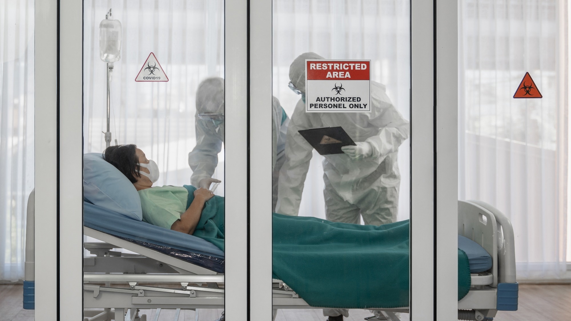
In a recent study posted to the bioRxiv* preprint server, researchers investigated the expression of complement regulatory proteins (CRPs) in coronavirus disease 2019 (COVID-19) to identify potential dysregulated CRP patterns and investigate whether they contribute to COVID-19 pathogenesis.
 Study: Upregulation of CD55 complement regulator in distinct PBMC subpopulations of COVID-19 patients is associated with suppression of interferon responses. Image Credit: Mongkolchon Akesin/Shutterstock
Background
Studies have reported complement activation in coronavirus disease 2019 (COVID-19) patients by elevated serological expression of complement factors C3a and C5b-9 and enhanced complement deposition in tissues. CRPs such as the cluster of differentiation (CD)46, CD55, CR1 (complement C3b/C4b receptor 1), and CD59 control complement overactivation and prevent complement deposition and lysis of cells.
However, data on the role of CRPs during viral infections through their complement cascade activation regulation, potential direct effects on antiviral processes, and immunological responses are lacking.
About the study
In the present study, researchers evaluated interferon (IFN)-stimulated gene (ISG) levels and CRP levels in severe acute respiratory syndrome coronavirus 2 (SARS-CoV-2) infections to examine dysregulated CRP patterns in COVID-19 and their contribution in complement overactivation.
Single-cell ribonucleic acid (RNA)-sequencing (scRNA-seq, with 61,022 single cells) analysis was performed on peripheral blood mononuclear cells (PBMCs) isolated from hospitalized SARS-CoV-2-infected patients to characterize immune responses, assess CRP expression signatures in different cell populations and explore the potential CRP involvement in COVID-19 pathogenesis.
Healthy individuals without COVID-19 served as controls, from whom, 15,705 single cells were subjected to scRNA-seq analysis. An integrated sub-clustering analysis of monocytes, T lymphocytes and B lymphocytes was performed to explore CRP expression in particular cell subpopulations.
In addition, PBMC subpopulation discriminatory analyses, flow cytometry (FC) analysis, functional gene ontology (GO), gene set enrichment analysis (GSEA) and UMAP analysis were performed. CD55 and CD59 expression patterns in monocytes, B and T lymphocytes of COVID-19 patients were assessed using FACS (fluorescence-activated cell sorting) analysis. ISGs such as IFN alpha inducible protein (IFI)- 6, 27, TM3, 44L, T1, T3, STAT2 (signal transducer and activator of transcription 2), ISG-15 and 20 expressions were assessed in B and T lymphocytes and monocytes.
Results and discussion
The scRNA-seq analysis, integrated sub-clustering analysis and FACS analysis showed CD55 and CD59 upregulation among individuals with severe and critical COVID-19 than in controls in monocytes and T lymphocytes. The FC analysis confirmed the distinctive pattern of elevated CD55 levels in monocytes and T lymphocyte sub-populations among individuals with severe COVID-19. CD55 upregulation was associated with lower ISG levels in severe COVID-19 for the CD14+ IL1B classical monocyte cluster, CD14+CD16+C-X-C motif chemokine ligand 10 (CXCL10) intermediate monocyte cluster, and the CD16+ non-classical monocyte cluster.
CD55 levels were higher in CD14+ monocytes than in CD16+ monocytes. Expression of IFI-6, ISG-15, and ISG-20 was the greatest among all COVID-19 patients and control individuals and was maintained among those with severe SARS-CoV-2 infections, contrasting ISG expression among monocytes wherein all ISGs showed similar expression patterns. ISG expression in T lymphocytes of critically ill COVID-19 patients were comparable to those in controls. Elevated CD55 expression was observed among CD4+ T lymphocytes than CD8+ lymphocytes and natural killer (NK) cells, indicating ISG reduction may be associated with CD55 upregulation among CD4+ T cells.
Elevated CD55 levels in severe SARS-CoV-2 infection T lymphocyte clusters were significant for most of the clusters except for the CD8+ cytotoxic exhausted cell cluster. Lower IFN responses were associated with higher CD55 levels in B lymphocytes. Similar trends of lower ISG levels in severe COVID-19 and critically ill COVID-19 post-elevation in mild SARS-CoV-2 infections were observed. ISG levels were identical to those noted among T lymphocytes, with the greatest expression of ISG-15 and 20 and sustained ISG-20 expression in severe SARS-CoV-2 infections.
Lower ISG expression was associated with higher CD55 expression among all B lymphocyte clusters of individuals with severe SARS-CoV-2 infections. Expression of IFI-6, 27, TM3, 44L, T1 and T3 ISGs increased to levels observed in controls and were nearly undetectable. Contrastingly, STAT2, ISG-15, and 20 showed sustained higher expressions among all cellular clusters, including those for individuals with critical SARS-CoV-2 infections. Plasmablasts and plasma cells showed the greatest expression of ISG-20 and 15 in stark contrast to other cellular clusters, respectively.
The elevation in CD55 levels was obvious among all severely SARS-CoV-2-infected patient clusters compared to controls, although statistical significance was not attained. CD46 was upregulated in monocytes, T and B lymphocytes. Of interest, reduced CD55 expression was observed in monocytes of patients with critical COVID-19 compared to severe SARS-CoV-2 infection patients, which may contribute to the sustained complement deposition in COVID-19 convalescent monocytes.
Increased CD59 expression in monocytes may have occurred since CD59 acts at the final step of the cascade, inhibiting C5b-9 formation and deposition and thus complement-mediated cell lysis, in monocytes and granulocytes of severe and critically ill SARS-CoV-2 infection patients. CD55 and CD46 upregulation in severe and critically ill COVID-19 patients may be an extra effort to activate the complement activation halting machinery and protect PBMCs that form the initial line of defense.
Overall, the study findings highlighted COVID-19-specific CD55 upregulation in PBMCs associated with lower IFN responses, indicating a potential role of CD55 in the pathogenesis of SARS-CoV-2 infections.
*Important notice
bioRxiv publishes preliminary scientific reports that are not peer-reviewed and, therefore, should not be regarded as conclusive, guide clinical practice/health-related behavior, or treated as established information.
- M. G. Detsika et al. (2022). Upregulation of CD55 complement regulator in distinct PBMC subpopulations of COVID-19 patients is associated with suppression of interferon responses. bioRxiv. doi:https://doi.org/10.1101/2022.10.07.510750 https://www.biorxiv.org/content/10.1101/2022.10.07.510750v1
Posted in: Medical Science News | Medical Research News | Disease/Infection News
Tags: B Lymphocyte, Blood, CD4, Cell, Cell Lysis, Cell Sorting, Chemokine, Coronavirus, Coronavirus Disease COVID-19, covid-19, CXCL10, Cytometry, Flow Cytometry, Fluorescence, Gene, Genes, Interferon, Ligand, Lymphocyte, Monocyte, Protein, Receptor, Respiratory, Ribonucleic Acid, RNA, SARS, SARS-CoV-2, Severe Acute Respiratory, Severe Acute Respiratory Syndrome, Syndrome, T Lymphocyte, Transcription

Written by
Pooja Toshniwal Paharia
Dr. based clinical-radiological diagnosis and management of oral lesions and conditions and associated maxillofacial disorders.
Source: Read Full Article
