
The term biotinylation refers to the process of binding biotin to either a protein or a nucleic acid, or in some cases to another type of molecule.
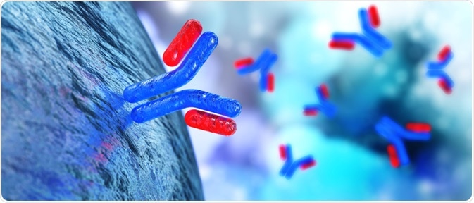
Image Credit: Utas7777777/Shutterstock.com
An antibody is one of these potential targets, a blood protein that responds to the presence of specific antigens that can be used as probes in cell research. In short, a biotinylated antibody is one of these blood proteins with biotin attached to it.
There are two main methods of antibody biotinylation, the avidin-biotin complex (ABC) method, and the labeled streptavidin-biotin (LSAB) method, both of which are discussed in detail.
How biotinylated antibodies are created
Biotin is a substance found naturally in different food types, such as eggs, dairy products, salmon, liver, and many more. Also known as vitamin H or vitamin B₇, biotin is essential to many key biological processes within the human body, mostly metabolic processes related to the utilization of amino acids, carbohydrates, and fats.
The process of biotinylation is fast, and because the molecule is relatively small, its attachment to an antibody is not thought to significantly impact its normal functioning. There is no one single accepted protocol for the biotinylation of antibodies.
However, the basis of the model relies on the strong interaction between biotin and avidin or streptavidin, which are considered to be some of the strongest non-covalent interactions between a protein and ligand.
These interactions are also immune to the pressures of pH and temperature extremes, as well as high salt concentrations and denaturants, making the biotin-avidin/streptavidin interaction incredibly useful for targeting proteins of interest.
A standard method of antibody biotinylation involves using N-Hydroxysulfosuccinimide (NHS) esters of biotin as the biotinylation reagent. NHS is the most popular type of reagent used, although it is possible to complete the biotinylation process with other kinds of reagents.
NHS (or a substitute) is used to activate the biotin, which forms stable amide bonds with primary amino acid groups within an alkaline buffer. The sidechain of lysine (K) residues and the N-terminus of each polypeptide are primary amines that act as common labeling targets on the antibody.
Avidin vs streptavidin methods
The basic method of antibody biotinylation can be split into two separate techniques relating to whether avidin or streptavidin interactions are utilized. If avidin is used, the avidin-biotin complex (ABC) method is appropriate.
This is where avidin-biotin complexes connected through reporter enzymes are incubated with biotinylated antibodies. The primary antibody is first incubated with the sample to allow for binding with the target antigen.
Following this, a biotinylated secondary antibody is incubated along with the tissue, causing binding to the primary antibody. A biotinylated enzyme is then pre-incubated with free avidin, resulting in avidin-biotin-enzyme complexes.
A portion of this mixture is then added to the sample, where remaining biotin-binding sites on the avidin bind to the biotinylated antibody that is already attached to the tissue sample. The sensitivity of this method can be raised by increasing the number of enzyme molecules that bind to the antigen, which can be achieved through increasing localized enzyme activity through reporter intensity.
The second method, the labeled streptavidin-biotin (LSAB) method, involves amplification of the signal through incubating the biotinylated antibody/streptavidin complexes along with reporter enzymes. The complexes created in the LSAB method are smaller than the above-described ABC method. In this method, streptavidin–enzyme conjugates are utilized for the detection of bound biotinylated primary antibodies on tissue samples. In most cases, the ABC method will be the first consideration unless the avidin-biotin-enzyme complexes become too large and can no longer pass through the tissue.
The LSAB staining method generates smaller complexes that can easily infiltrate the tissue, having the impact of improving sensitivity by up to eight times that of the ABC method. Alternatives to avidin can be used in this method, which can have the effect of reducing background noise and further increasing sensitivity.
The LSAB method involves two main stages. First, the primary antibodies are incubated alongside the biotinylated secondary antibodies, with the tissue sample using the same process outlined in the ABC method. Following this, enzyme-conjugated streptavidin is added and incubated, resulting in the biotin-binding sites on the enzyme-conjugated protein becoming occupied.
Functions and limitations of biotinylated antibodies
Biotinylated antibodies are used for the detection of low-abundance proteins. The process of biotin-labeling is also frequently used as a non-radiative labeling method of proteins, and as a protein purification technique.
While there are many benefits to this method, such as its non-radioactive status, which has led to its wide adoption, it also has a notable limitation.
As biotin is used by the body for many processes, it is natural that endogenous biotin in tissues may be present, having the effect of increasing the background signal. Because of this, measures to block endogenous biotin are required when investigating tissues with high biotin expressions, such as those from the liver or kidney.
Sources:
Bratthauer, G. (2009). The Avidin-Biotin Complex (ABC) Method and Other Avidin-Biotin Binding Methods. Immunocytochemical Methods and Protocols, pp.257-270. https://www.ncbi.nlm.nih.gov/pubmed/20012837
Mao, S. (2009). Biotinylation of Antibodies. Immunocytochemical Methods and Protocols, pp.49-52. https://link.springer.com/protocol/10.1007%2F978-1-59745-324-0_7
Strachan, E., Mallia, A., Cox, J., Antharavally, B., Desai, S., Sykaluk, L., O'Sullivan, V. and Bell, P. (2004). Solid-phase biotinylation of antibodies. Journal of Molecular Recognition, 17(3), pp.268-276. https://www.ncbi.nlm.nih.gov/pubmed/15137036
Further Reading
- All Antibody Content
- Antibody – What is an Antibody?
- Antibody Forms
- Antibodies in Medicine
- Antibody Structure
Last Updated: Jan 30, 2020

Written by
Sarah Moore
After studying Psychology and then Neuroscience, Sarah quickly found her enjoyment for researching and writing research papers; turning to a passion to connect ideas with people through writing.
Source: Read Full Article