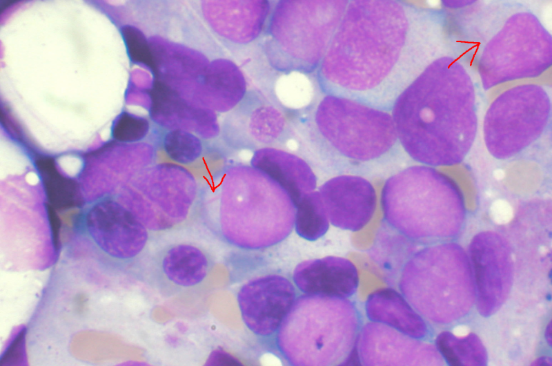
Myelodysplastic syndrome (MDS) is a disease of the stem cells in the bone marrow, which disturbs the maturing and differentiation of blood cells. Annually, some 200 Finns are diagnosed with MDS, which can develop into acute leukemia. Globally, the incidence of MDS is 4 cases per 100,000 person years.
To diagnose MDS, a bone marrow sample is needed to also investigate genetic changes in bone marrow cells. The syndrome is classified into groups to determine the nature of the disorder in more detail.
In the study conducted at the University of Helsinki, microscopic images of MDS patients’ bone marrow samples were examined utilizing an image analysis technique based on machine learning. The samples were stained with haematoxylin and eosin (H&E staining), a procedure that is part of the routine diagnostics for the disease. The slides were digitized and analyzed with the help of computational deep learning models.
The study was published in the Blood Cancer Discovery, a journal of the American Association for Cancer Research, and the results can also be explored with an interactive tool: hruh-20.it.helsinki.fi/mds_visualization/ .
By employing machine learning, the digital image dataset could be analyzed to accurately identify the most common genetic mutations affecting the progression of the syndrome, such as acquired mutations and chromosomal aberrations. The higher the number of aberrant cells in the samples, the higher the reliability of the results generated by the prognostic models.
Diagnosis supported by data analysis
One of the greatest challenges of utilizing neural network models is understanding the criteria on which they base their conclusions drawn from data, such as information contained in images. The recently released study succeeded in determining what deep learning models see in tissue samples when they have been taught to look for, for example, genetic mutations related to MDS. The technique provides new information on the effects of complex diseases on bone marrow cells and the surrounding tissues.
“The study confirms that computational analysis helps to identify features that elude the human eye. Moreover, data analysis helps to collect quantitative data on cellular changes and their relevance to the patient’s prognosis,” says Professor Satu Mustjoki.
Part of the analytics carried out in the study was implemented using the Helsinki University Hospital (HUS) data lake environment, which enables the efficient collection and analysis of extensive clinical datasets.
“We’ve developed solutions to structure and analyze data stored in the HUS data lake. Image analysis helps us analyze large quantities of biopsies and rapidly produce diverse information on disease progression. The techniques developed in the project are suited to other projects as well, and they are perfect examples of the digitalizing medical science,” says doctoral student Oscar Bruck.
Source: Read Full Article
