The gut-brain connection is a complicated two-way signaling cascade that is responsible for keeping the digestive system working properly and can cause problems when it breaks down. A key part of that axis is the colon, which extracts water and nutrients from food and transports waste out of the body. This crucial organ is implicated in a range of gastrointestinal conditions, including constipation, diarrhea, pain, and inflammation.
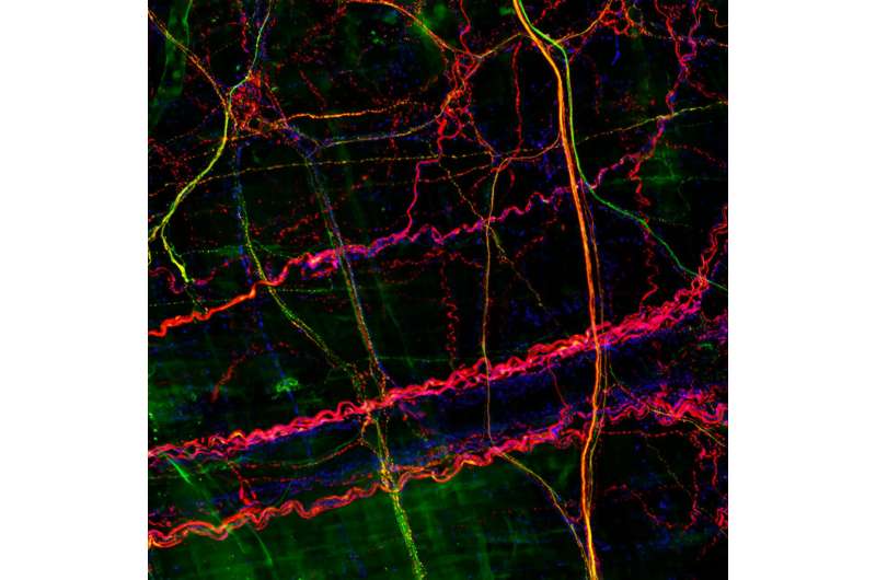
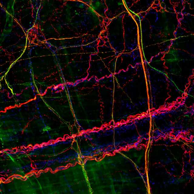
Now, in a first, researchers at Harvard Medical School have defined five distinct subtypes of sensory neurons in the colon that carry signals to the brain.
In a new study, conducted in mice and published Aug. 3 in Cell, the researchers found that some neurons are dedicated to sensing gentle forces, such as substances moving through the colon, while others sense more intense ones, such as pain.
The researchers say that, if confirmed in humans, their findings could help scientists develop more effective therapies to treat conditions that arise when this colon-brain sensing system goes awry.
“Patients often complain about sensation and pain in the gastrointestinal system, yet we don’t know a lot about the sensory neurons that innervate the gut and allow us to respond to different stimuli,” said lead author Rachel Wolfson, a research fellow in neurobiology at HMS and a gastroenterology fellow at Massachusetts General Hospital.
From skin to colon
David Ginty and scientists in his lab have spent many years studying how sensory neurons in the skin communicate with the brain to form our sense of touch. They have developed precise genetic tools that label subtypes of sensory neurons and used these tools to uncover basic information about the structure, organization, and function of skin-sensing neurons.
Yet even as scientific knowledge about touch neurons has grown, very little research has focused on understanding the neurons in other parts of the body, including the gastrointestinal system.
“We’ve learned a lot about the neurons that go to the skin, but the properties of neurons that project to other organs like the colon have remained poorly understood,” said Ginty, the Edward R. and Anne G. Lefler Professor of Neurobiology in the Blavatnik Institute at HMS and senior author on the new paper.
To tackle this understudied area, Ginty teamed up with Wolfson, a neurobiologist and clinical expert on the gastrointestinal system.
Wolfson took genetically-labeled mouse models developed in the Ginty lab and repurposed them to study neurons in the colon. She discovered that five subtypes of sensory neurons in the skin are also found in the colon. However, colon and touch neurons had distinct shapes, and the subtypes of colon neurons also varied from each other in form.
“We know that form underlies function, so the fact that the colon neurons look different from each other made us think that they have different functions,” Wolfson said.
To investigate function, Wolfson stretched the colon with a balloon—mimicking naturally occurring distension—and recorded activity in the distinct types of neurons.
Two types responded to gentle forces, similar to the slight stretching that might happen when digested food or stool moves through the colon. Two other types responded to intense forces, such as more extreme stretching. When Wolfson artificially activated these high-force neurons, the mice behaved as if they were in pain. When she removed the neuron with the highest force threshold, the pain response diminished. Triggering inflammation in the mice caused one of the pain-sensing neuron subtypes to become even more reactive.
Interestingly, these roles mapped onto the roles of neurons in the skin, suggesting that function may be conserved across organ systems.
The study provides critical insight into the basic neurobiological mechanisms of colon sensation.
“For the first time, we’ve been able to figure out the anatomy, physiology, and functions of neurons that innervate the colon,” Ginty said.
Toward therapies for GI problems
In the short term, the researchers want to understand why colon neurons look different from their skin counterparts, and how these differences in form translate into differences in the way they behave.
“This finding is really provocative and provides a whole other direction for the work around understanding how the neurons convert mechanical forces into electrical signals, which is the currency of the nervous system,” Ginty said.
In the longer term, Wolfson plans to study the neurons in other parts of the gastrointestinal tract. She also wants to explore how colon neurons respond to other stimuli such as toxins or a lack of blood flow, which can cause abdominal pain.
While the results would first have to be confirmed in humans, the researchers say their work could one day inform the development of better therapies for various gastrointestinal conditions.
“We’ve known about the innervation of the gut for 100 years, but modern neuroscience tools allow us to dig in and understand how it all works, and that’s going to serve as a platform for therapeutic approaches to treating colon problems,” Ginty said.
Targeting low-force neurons could be helpful for treating motility-related conditions like constipation and diarrhea, while targeting high-force neurons could be useful for treating pain that originates in the colon. Of particular interest is the neuron subtype that is sensitive to inflammation, which is a source of pain for patients with inflammatory bowel disease.
“Having a way to target these neurons to treat a patient’s pain while we’re getting the inflammation under control with anti-inflammatory medications is a huge therapeutic need,” Wolfson said.
Additional authors on the paper include Amira Abdelaziz, Genelle Rankin, Sarah Kushner, Lijun Qi, Ofer Mazor, and Seungwon Choi of HMS and Nikhil Sharma of HMS and Columbia University.
More information:
David D. Ginty, DRG afferents that mediate physiologic and pathologic mechanosensation from the distal colon, Cell (2023). DOI: 10.1016/j.cell.2023.07.007. www.cell.com/cell/fulltext/S0092-8674(23)00740-7
Journal information:
Cell
Source: Read Full Article
