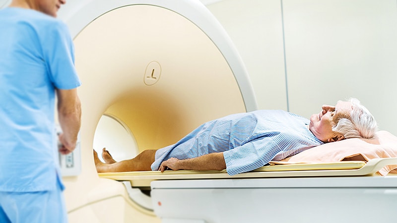
Including cardiac CT in the initial hyperacute stroke imaging protocol gives a higher yield for detecting high-risk cardioaortic sources of embolism than the current practice of performing later echocardiography, a new study has shown.
“We found an earlier detection and higher yield of cardiac embolism by incorporating cardiac CT into the regular CT imaging already routinely carried out in acute stroke patients,” senior author, Jonathan M. Coutinho, MD, University of Amsterdam, Amsterdam, Netherlands, commented to theheart.org | Medscape Cardiology.
“We also found that adding in a cardiac CT scan is feasible. Patients are already in the scanner. We are doing other CT scans anyway, so adding in one extra scan is very straightforward and quick, extending the total scan protocol by only 6 minutes,” he said.
“Neurologists need to start taking notice of cardiac CT. Although this is the first prospective study comparing the two techniques, I think we are on a trajectory where cardiac CT may end up replacing echocardiography as a first line screening method in stroke patients,” Coutinho added.
The study was published in the Oct. 4 issue of Neurology.
Results showed that a high-risk cardioaortic source of embolism was detected five times more frequently with early cardiac CT than with later echocardiography.
Commenting in a podcast, available on the Neurology website, Daniel Ackerman, MD, director of Stroke and Vascular Neurology at St. Luke’s University Health Network, Pennsylvania, described the results as “amazing.”
Noting that up to one third of strokes remain cryptogenic even after all the routine tests and imaging are conducted, leading to the term “embolic stroke of undetermined origin (ESUS),” Ackerman said, adding, “This is a new and exciting way of evaluating stroke patients so that this can be translated to ’embolic stroke of determined origin.'”
And authors of an accompanying editorial, with the strapline “Don’t Leave the CT Scanner Without Imaging the Heart,” describe the study as “landmark,” and conclude that: “The role of the CT scanner for the hyperacute stroke workup may soon not only become a ‘one-stop shop’ for acute reperfusion therapy but also guide a new paradigm for early secondary stroke prevention in cardioembolic stroke.”
The “Mind the Heart” study included 452 consecutive patients who underwent ECG-gated cardiac CT in the hyperacute stroke phase. Of these, 350 also underwent transthoracic echocardiography at a later timepoint (median, 1 day later).
In the group who had both imaging modalities, a high-risk cardioaortic embolic source was detected in 11.4% with cardiac CT compared with only 4.9% on transthoracic echocardiography (odds ratio, 5.60; 95% CI, 2.28-16.33).
The diagnostic yield of cardiac CT in the full study population, including patients who were unable to undergo echocardiography was 12.2%.
Intracardiac thrombus was the most frequent finding, and it was identified considerably more often with cardiac CT, 7.1%, vs 0.6% on echocardiography.
Among the 175 patients with cryptogenic stroke after the stroke workup, cardiac CT identified a cause of the stroke in 6.3%.
Coutinho explained that transthoracic echocardiography is the traditional method of imaging to search for cardioembolism after an acute ischemic stroke. Often this is not done in the hyperacute phase but in the days or weeks following the stroke.
“However, this imaging has a low yield and we always felt it is not fulfilling our needs,” he said.
“We need to find the smoking gun — the actual cause of the stroke — and we thought the chances of picking up a thrombus would be higher if imaging is conducted sooner after the stroke occurs, preferably even before thrombolysis is given, as thrombolysis may dissolve the thrombus,” Coutinho noted.
“We wanted to look at conducting a chest CT to look for cardioembolism at the same time that the patient has routine head CTs when first presenting with a suspected stroke. This can be done in the same machine in many cases. While it may depend on what type of CT scanner is being used, most modern CT scanners will be able to do this.”
The researchers used ECG-gated CT in which the scan is triggered by a specific phase of the ECG. While this takes a few minutes longer than non-gated cardiac CT, as the patient has to be repositioned, it produces higher-quality images.
In the current study, the gated CT scan added 6 minutes to the imaging protocol.
Coutinho does not believe adding this cardiac CT scan in the hyperacute phase will delay time to thrombolysis.
“We always say starting thrombolysis should be prioritized over doing the cardiac CT scan, but in reality, that hardly ever happens, as it usually takes a few minutes for the regular CT scans to be evaluated before deciding on thrombolysis and during this time the cardiac CT can be done. But this needs to be carefully planned when incorporating cardiac CT into stroke protocols,” he said.
He also pointed out that a cardiac CT scan in the hyperacute phase also has other potential benefits, such as picking up other conditions that could affect management decisions. For example, if endocarditis is seen, then thrombolysis may not be indicated because of an increased risk of hemorrhage.
There were eight patients in the study in whom a high-risk source of embolism was detected with echocardiography that was not identified on CT, and these findings changed patient management in two patients (one who had signs of endocarditis and one who had signs of a recent myocardial infarction).
Addressing this, Coutinho said, “We need to figure out for which patients cardiac echo is still indicated. I think we can learn a lot here from taking a good history and clinical examination. Most patients with endocarditis will have some clinical signs of infection as the cause of stroke.”
He added that recognizing a patent foramen ovale (PFO) on CT may be more difficult than echo, “so perhaps for young patients with cryptogenic stroke in whom we may consider closing a PFO, an echo may still be indicated.”
The authors of the editorial, Mark W. Parsons, PhD, University of New South Wales, Sydney, Australia, and Carlos Garcia-Esperon, PhD, University of Newcastle, Callaghan, Australia, raise some further questions on the study.
Noting that there was a relatively high proportion of patients with large vessel occlusion stroke included, they wonder if the results would be applicable to a milder cohort. And they ask whether a more rapidly acquired non-gated cardiac CT would also detect the same rates of atrial thrombus.
Study funding included grants from the Royal Netherlands Academy of Arts and Sciences, Foundation De Drie Lichten, Remmert Adriaan Laan Foundation, and AMC Young Talent Fund, all of which are nonprofit research foundations. Dr Coutinho reports grants from Medtronic (paid to institution).
Neurology. Oct. 4 issue. Abstract; Editorial.
For more from theheart.org | Medscape Cardiology, follow us on Twitter and Facebook.
Source: Read Full Article
