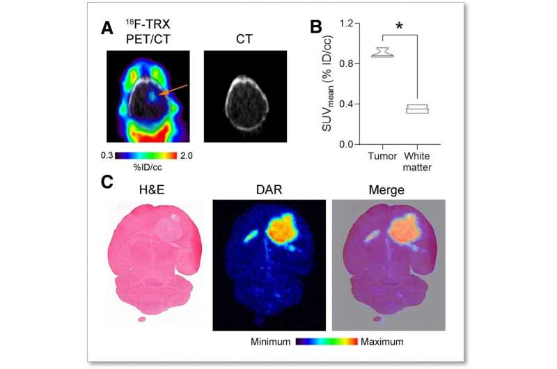
A new radiotracer that detects iron in cancer cells has proven effective, opening the door for the advancement of iron-targeted therapies for cancer patients. The radiotracer, 18F-TRX, can be used to measure iron concentration in tumors, which can help predict whether a not the cancer will respond to treatment. This research was published in the July issue of the Journal of Nuclear Medicine.
All cancer cells have an insatiable appetite for iron, which provides them the energy they need to multiply. As a result, tumors have higher levels of iron than normal tissues. Recent advances in chemistry have led scientists to take advantage of this altered state, targeting the expanded cytosolic ‘labile’ iron pool (LIP) of the cancer cell to develop new treatments.
A clear method to measure LIP in tumors must be established to advance clinical trials for LIP-targeted therapies. “LIP levels in patient tumors have never been quantified,” noted Adam R. Renslo, Ph.D., professor in the department of pharmaceutical chemistry at the University of California, San Francisco. “Iron rapidly oxidizes once its cellular environment is disrupted, so it can’t be quantified reliably from tumor biopsies. A biomarker for LIP could help determine which tumors have the highest LIP levels and might be especially vulnerable to LIP-targeted therapies.”
To explore a solution for this unmet need, researchers imaged 10 tissue graft models of glioma and renal cell carcinoma with 18F-TRX PET to measure LIP. Tumor avidity and sensitivity to the radiotracer were assessed. An animal model study was also conducted to determine effective human dosimetry.
18F-TRX showed a wide range of tumor accumulation, successfully distinguishing LIP levels among tumors and determining those that might be most likely to respond to LIP-targeted therapies. Pretreatment 18F-TRX uptake in tumors was also found to predict sensitivity to therapy. The estimated effective dose for adults was comparable to those of other 18F-based imaging agents.
Source: Read Full Article
